All organs and systems of the human body are closely interconnected, they interact with the help of the nervous system, which regulates all the mechanisms of life, from digestion to the process of reproduction. It is known that a person (NS) provides a connection between the human body and the external environment. The unit of the NS is the neuron, which is a nerve cell that conducts impulses to other cells of the body. Connecting into neural circuits, they form a whole system, both somatic and vegetative.
We can say that the NS is plastic, as it is able to restructure its work in the case when the needs of the human body change. This mechanism is especially relevant when one of the parts of the brain is damaged.
Since the human nervous system coordinates the work of all organs, its damage affects the activity of both nearby and distant structures, and is accompanied by the failure of the functions of organs, tissues and body systems. The causes of disruption of the nervous system may lie in the presence of infections or poisoning of the body, in the occurrence of a tumor or injury, in diseases of the National Assembly and metabolic disorders.
Thus, the human NS plays a conducting role in the formation and development of the human body. Thanks to the evolutionary improvement of the nervous system, the human psyche and consciousness developed. The nervous system is a vital mechanism for regulating the processes that occur in the human body.
The human nervous system is made up of tiny cells called nerve cells. Through circuits made up of these cells, nerve impulses go to the brain, and the response - to the muscles. There are over 10 billion nerve cells in the human body.
Different areas of the brain are responsible for a variety of feelings, sensations and moods.
Nerve cells are called neurons. Outwardly, neurons have a variety of shapes: some are star-shaped, others are triangles or spirals. But even such a small detail of the body as neuron, consists of several parts: body, long process - axon and shorter and thinner processes - dendrites. Thanks to the processes, the cells are attached to each other and their interaction. The body of a neuron, like any other cell, consists of a nucleus surrounded by cytoplasm and covered with a membrane.
The central organ of the human nervous system that controls its functioning is brain. The human brain is able to perform much more processes associated with thinking, feelings, emotions than the brain of other living beings. The surface of the human brain is covered with shallow furrows - convolutions. It consists of white and gray matter. With the help of the first there is a connection between the spinal cord and the brain, and the second makes up the cerebral cortex.

The human brain is made up of several sections.
medulla oblongata and pons serve to communicate with the spinal cord. They control the work of the digestive and respiratory systems, the work of the heart.
Cerebellum coordinates all human movements. It is the activity of this part of the brain that ensures the accuracy and speed of movements.
midbrain responsible for the reaction to external stimuli, that is, responsible for the sensory system.
diencephalon regulates metabolism and body temperature.
The largest parts of the brain are two cerebral hemispheres. The hemispheres of the brain allow a person to analyze the sensations received through the senses (for example, the taste of food). The hemispheres of the brain are also responsible for speech, thinking, emotions.
brain weight- on average, it is 1360-1375 grams for men, 1220-1245 grams for women. After rapid growth during the first year of life (the brain of a newborn is 410 grams - 1/8 of the body weight; the brain weight at the end of the first year is 900 grams - 1/14 of the body weight), the brain grows slowly and between 20-30 years reaches the limit of its growth, up to 50 years does not change, and then begins to decrease in weight. Among animals, man has the greatest weight of the brain, not only relative, but also absolute. Only the whale has a slightly heavier brain than a human (2816). The brain of a horse weighs 680 g; lion - 250 g; anthropomorphic monkeys 350-400, rarely more.
The greater or lesser weight of the brain in different people cannot in itself be an indication of the size of their mental abilities. On the other hand, people of outstanding ability often have a brain weight that far exceeds the average. The richness of mental organization depends on the quantity and quality of nerve cells in the cortical layer of the hemispheres and, probably, on the number of association fibers of the large brain.
The second most important organ of the nervous system is spinal cord. It is located inside the dorsal and cervical vertebrae. The spinal cord is responsible for all human movements and is connected to the brain, which coordinates these movements. The spinal cord together with the brain make up the central nervous system, and the nerve processes make up the peripheral nervous system.
LECTURE ON THE TOPIC: HUMAN NERVOUS SYSTEM
Nervous system is a system that regulates the activity of all human organs and systems. This system determines: 1) the functional unity of all human organs and systems; 2) the connection of the whole organism with the environment.
From the point of view of maintaining homeostasis, the nervous system provides: maintaining the parameters of the internal environment at a given level; inclusion of behavioral responses; adaptation to new conditions if they persist for a long time.
Neuron(nerve cell) - the main structural and functional element of the nervous system; Humans have over 100 billion neurons. The neuron consists of a body and processes, usually one long process - an axon and several short branched processes - dendrites. Along the dendrites, impulses follow to the cell body, along the axon - from the cell body to other neurons, muscles or glands. Thanks to the processes, neurons contact each other and form neural networks and circles through which nerve impulses circulate.
A neuron is the functional unit of the nervous system. Neurons are susceptible to stimulation, that is, they are able to be excited and transmit electrical impulses from receptors to effectors. In the direction of impulse transmission, afferent neurons (sensory neurons), efferent neurons (motor neurons) and intercalary neurons are distinguished.
Nervous tissue is called excitable tissue. In response to some impact, the process of excitation arises and spreads in it - the rapid recharging of cell membranes. The emergence and spread of excitation (nerve impulse) is the main way the nervous system implements its control function.
The main prerequisites for the occurrence of excitation in cells: the existence of an electrical signal on the membrane at rest - the resting membrane potential (RMP);
the ability to change the potential by changing the permeability of the membrane for certain ions.
The cell membrane is a semi-permeable biological membrane, it has channels for potassium ions to pass through, but there are no channels for intracellular anions that are held at the inner surface of the membrane, while creating a negative charge of the membrane from the inside, this is the resting membrane potential, which is on average - - 70 millivolts (mV). There are 20-50 times more potassium ions in the cell than outside, this is maintained throughout life with the help of membrane pumps (large protein molecules capable of transporting potassium ions from the extracellular environment to the inside). The MPP value is due to the transfer of potassium ions in two directions:
1. outside into the cage under the action of pumps (with a large expenditure of energy);
2. out of the cell by diffusion through membrane channels (without energy costs).
In the process of excitation, the main role is played by sodium ions, which are always 8-10 times more outside the cell than inside. Sodium channels are closed when the cell is at rest, in order to open them, it is necessary to act on the cell with an adequate stimulus. If the stimulation threshold is reached, sodium channels open and sodium enters the cell. In thousandths of a second, the membrane charge will first disappear, and then change to the opposite - this is the first phase of the action potential (AP) - depolarization. The channels close - the peak of the curve, then the charge is restored on both sides of the membrane (due to potassium channels) - the stage of repolarization. Excitation stops and while the cell is at rest, the pumps change the sodium that has entered the cell for the potassium that has left the cell.

AP evoked at any point of the nerve fiber itself becomes an irritant for neighboring sections of the membrane, causing AP in them, and they, in turn, excite more and more new sections of the membrane, thus spreading throughout the cell. In myelin-coated fibers, PD will only occur in myelin-free areas. Therefore, the speed of signal propagation increases.


The transfer of excitation from a cell to another occurs with the help of a chemical synapse, which is represented by the point of contact between two cells. The synapse is formed by the presynaptic and postsynaptic membranes and the synaptic cleft between them. Excitation in the cell resulting from AP reaches the area of the presynaptic membrane, where synaptic vesicles are located, from which a special substance, the mediator, is ejected. The neurotransmitter enters the gap, moves to the postsynaptic membrane and binds to it. Pores for ions open in the membrane, they move inside the cell and a process of excitation occurs.

Thus, in the cell, the electrical signal is converted into a chemical one, and the chemical signal is again converted into an electrical one. Signal transmission in the synapse is slower than in the nerve cell, and also one-sided, since the mediator is released only through the presynaptic membrane, and can only bind to the receptors of the postsynaptic membrane, and not vice versa.
Mediators can cause in cells not only excitation, but also inhibition. At the same time, pores are opened on the membrane for such ions, which increase the negative charge that existed on the membrane at rest. One cell can have many synaptic contacts. An example of a mediator between a neuron and a skeletal muscle fiber is acetylcholine.
The nervous system is divided into central nervous system and peripheral nervous system.
In the central nervous system, the brain is distinguished, where the main nerve centers and the spinal cord are concentrated, here there are centers of a lower level and there are pathways to peripheral organs.
Peripheral - nerves, ganglia, ganglia and plexuses.
The main mechanism of activity of the nervous system - reflex. A reflex is any response of the body to a change in the external or internal environment, which is carried out with the participation of the central nervous system in response to irritation of the receptors. The structural basis of the reflex is the reflex arc. It includes five consecutive links:
1 - Receptor - a signaling device that perceives the impact;
2 - Afferent neuron - leads the signal from the receptor to the nerve center;
3 - Intercalary neuron - the central part of the arc;
4 - Efferent neuron - the signal comes from the central nervous system to the executive structure;
5 - Effector - a muscle or gland that performs a certain type of activity

Brain consists of accumulations of bodies of nerve cells, nerve tracts and blood vessels. Nerve tracts form the white matter of the brain and consist of bundles of nerve fibers that conduct impulses to or from various areas of the gray matter of the brain - the nuclei or centers. Pathways connect the various nuclei, as well as the brain with the spinal cord.
Functionally, the brain can be divided into several sections: the forebrain (consisting of the telencephalon and diencephalon), the midbrain, the hindbrain (consisting of the cerebellum and the pons), and the medulla oblongata. The medulla oblongata, pons, and midbrain are collectively referred to as the brainstem.

Spinal cord located in the spinal canal, reliably protecting it from mechanical damage.
The spinal cord has a segmental structure. Two pairs of anterior and posterior roots depart from each segment, which corresponds to one vertebra. There are 31 pairs of nerves in total.
The posterior roots are formed by sensitive (afferent) neurons, their bodies are located in the ganglia, and the axons enter the spinal cord.
The anterior roots are formed by axons of efferent (motor) neurons whose bodies lie in the spinal cord.
The spinal cord is conditionally divided into four sections - cervical, thoracic, lumbar and sacral. It closes a huge number of reflex arcs, which ensures the regulation of many body functions.
The gray central substance is nerve cells, the white one is nerve fibers.

The nervous system is divided into somatic and autonomic.
To somatic nervous system (from the Latin word "soma" - body) refers to the part of the nervous system (both cell bodies and their processes), which controls the activity of skeletal muscles (body) and sensory organs. This part of the nervous system is largely controlled by our consciousness. That is, we are able to bend or unbend an arm, a leg, and so on at will. However, we are unable to consciously stop perceiving, for example, sound signals.
Autonomic nervous a system (translated from Latin “vegetative” - vegetable) is a part of the nervous system (both the cell body and their processes) that controls the processes of metabolism, growth and reproduction of cells, that is, functions that are common to both animals and plants organisms. The autonomic nervous system controls, for example, the activity of internal organs and blood vessels.
The autonomic nervous system is practically not controlled by consciousness, that is, we are not able, at will, to remove the spasm of the gallbladder, stop cell division, stop intestinal activity, expand or narrow blood vessels
NERVOUS SYSTEM
a complex network of structures that permeates the entire body and provides self-regulation of its vital activity due to the ability to respond to external and internal influences (stimuli). The main functions of the nervous system are the receipt, storage and processing of information from the external and internal environment, the regulation and coordination of the activities of all organs and organ systems. In humans, as in all mammals, the nervous system includes three main components: 1) nerve cells (neurons); 2) glial cells associated with them, in particular neuroglial cells, as well as cells that form neurilemma; 3) connective tissue. Neurons provide the conduction of nerve impulses; neuroglia performs supporting, protective and trophic functions both in the brain and spinal cord, and neurilemma, which consists mainly of specialized, so-called. Schwann cells, participates in the formation of sheaths of peripheral nerve fibers; connective tissue supports and links together the various parts of the nervous system. The human nervous system is divided in different ways. Anatomically, it consists of the central nervous system (CNS) and the peripheral nervous system (PNS). The central nervous system includes the brain and spinal cord, and the PNS, which provides communication between the central nervous system and various parts of the body, includes cranial and spinal nerves, as well as nerve nodes (ganglia) and nerve plexuses that lie outside the spinal cord and brain.
Neuron. The structural and functional unit of the nervous system is a nerve cell - a neuron. It is estimated that there are more than 100 billion neurons in the human nervous system. A typical neuron consists of a body (i.e., a nuclear part) and processes, one usually non-branching process, an axon, and several branching ones, dendrites. The axon carries impulses from the cell body to the muscles, glands, or other neurons, while the dendrites carry them to the cell body. In a neuron, as in other cells, there is a nucleus and a number of tiny structures - organelles (see also CELL). These include the endoplasmic reticulum, ribosomes, Nissl bodies (tigroid), mitochondria, the Golgi complex, lysosomes, filaments (neurofilaments and microtubules).

Nerve impulse. If the stimulation of a neuron exceeds a certain threshold value, then a series of chemical and electrical changes occur at the point of stimulation, which spread throughout the neuron. Transmitted electrical changes are called nerve impulses. Unlike a simple electric discharge, which, due to the resistance of the neuron, will gradually weaken and be able to overcome only a short distance, a much slower "running" nerve impulse in the process of propagation is constantly restored (regenerates). The concentrations of ions (electrically charged atoms) - mainly sodium and potassium, as well as organic substances - outside the neuron and inside it are not the same, so the nerve cell at rest is negatively charged from the inside, and positively from the outside; as a result, a potential difference appears on the cell membrane (the so-called "resting potential" is approximately -70 millivolts). Any change that reduces the negative charge inside the cell and thereby the potential difference across the membrane is called depolarization. The plasma membrane surrounding a neuron is a complex formation consisting of lipids (fats), proteins and carbohydrates. It is practically impermeable to ions. But some of the protein molecules in the membrane form channels through which certain ions can pass. However, these channels, called ionic channels, are not always open, but, like gates, they can open and close. When a neuron is stimulated, some of the sodium (Na +) channels open at the point of stimulation, due to which sodium ions enter the cell. The influx of these positively charged ions reduces the negative charge of the inner surface of the membrane in the region of the channel, which leads to depolarization, which is accompanied by a sharp change in voltage and a discharge - a so-called. "action potential", i.e. nerve impulse. The sodium channels then close. In many neurons, depolarization also causes potassium (K+) channels to open, causing potassium ions to flow out of the cell. The loss of these positively charged ions again increases the negative charge on the inner surface of the membrane. The potassium channels then close. Other membrane proteins also begin to work - the so-called. potassium-sodium pumps that ensure the movement of Na + from the cell, and K + into the cell, which, along with the activity of potassium channels, restores the initial electrochemical state (resting potential) at the point of stimulation. Electrochemical changes at the point of stimulation cause depolarization at the adjacent point of the membrane, triggering the same cycle of changes in it. This process is constantly repeated, and at each new point where depolarization occurs, an impulse of the same magnitude is born as at the previous point. Thus, together with the renewed electrochemical cycle, the nerve impulse propagates along the neuron from point to point. Nerves, nerve fibers and ganglia. A nerve is a bundle of fibers, each of which functions independently of the others. The fibers in a nerve are organized into clusters surrounded by specialized connective tissue, which contains vessels that supply the nerve fibers with nutrients and oxygen and remove carbon dioxide and waste products. Nerve fibers along which impulses propagate from peripheral receptors to the central nervous system (afferent) are called sensitive or sensory. Fibers that transmit impulses from the central nervous system to muscles or glands (efferent) are called motor or motor. Most nerves are mixed and consist of both sensory and motor fibers. A ganglion (ganglion) is a cluster of neuron bodies in the peripheral nervous system. Axon fibers in the PNS are surrounded by a neurilemma - a sheath of Schwann cells that are located along the axon, like beads on a thread. A significant number of these axons are covered with an additional sheath of myelin (a protein-lipid complex); they are called myelinated (meaty). Fibers that are surrounded by neurilemma cells, but not covered with a myelin sheath, are called unmyelinated (non-myelinated). Myelinated fibers are found only in vertebrates. The myelin sheath is formed from the plasma membrane of the Schwann cells, which winds around the axon like a roll of ribbon, forming layer upon layer. The area of the axon where two adjacent Schwann cells touch each other is called the node of Ranvier. In the CNS, the myelin sheath of nerve fibers is formed by a special type of glial cells - oligodendroglia. Each of these cells forms the myelin sheath of several axons at once. Unmyelinated fibers in the CNS lack a sheath of any special cells. The myelin sheath speeds up the conduction of nerve impulses that "jump" from one node of Ranvier to another, using this sheath as a connecting electrical cable. The speed of impulse conduction increases with the thickening of the myelin sheath and ranges from 2 m / s (along unmyelinated fibers) to 120 m / s (along fibers, especially rich in myelin). For comparison: the speed of propagation of electric current through metal wires is from 300 to 3000 km / s.
Synapse. Each neuron has a specialized connection to muscles, glands, or other neurons. The zone of functional contact between two neurons is called a synapse. Interneuronal synapses are formed between different parts of two nerve cells: between an axon and a dendrite, between an axon and a cell body, between a dendrite and a dendrite, between an axon and an axon. A neuron that sends an impulse to a synapse is called presynaptic; the neuron receiving the impulse is postsynaptic. The synaptic space is slit-shaped. A nerve impulse propagating along the membrane of a presynaptic neuron reaches the synapse and stimulates the release of a special substance - a neurotransmitter - into a narrow synaptic cleft. Neurotransmitter molecules diffuse through the cleft and bind to receptors on the membrane of the postsynaptic neuron. If the neurotransmitter stimulates the postsynaptic neuron, its action is called excitatory; if it suppresses, it is called inhibitory. The result of the summation of hundreds and thousands of excitatory and inhibitory impulses simultaneously flowing to a neuron is the main factor determining whether this postsynaptic neuron will generate a nerve impulse at a given moment. In a number of animals (for example, in the spiny lobster), a particularly close connection is established between the neurons of certain nerves with the formation of either an unusually narrow synapse, the so-called. gap junction, or, if neurons are in direct contact with each other, tight junction. Nerve impulses pass through these connections not with the participation of a neurotransmitter, but directly, by electrical transmission. A few dense junctions of neurons are also found in mammals, including humans.
Regeneration. By the time a person is born, all of his neurons and most of the interneuronal connections have already been formed, and in the future only single new neurons are formed. When a neuron dies, it is not replaced by a new one. However, the remaining ones can take over the functions of the lost cell, forming new processes that form synapses with those neurons, muscles or glands with which the lost neuron was connected. Cut or damaged PNS neuron fibers surrounded by neurilemma can regenerate if the cell body remains intact. Below the site of transection, the neurilemma is preserved as a tubular structure, and that part of the axon that remains connected with the cell body grows along this tube until it reaches the nerve ending. Thus, the function of the damaged neuron is restored. Axons in the CNS that are not surrounded by a neurilemma are apparently unable to grow back to the site of their former termination. However, many CNS neurons can give rise to new short processes - branches of axons and dendrites that form new synapses.
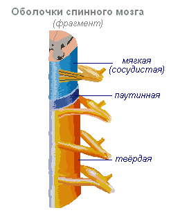
CENTRAL NERVOUS SYSTEM

The CNS consists of the brain and spinal cord and their protective membranes. The outermost is the dura mater, under it is the arachnoid (arachnoid), and then the pia mater, fused with the surface of the brain. Between the soft and arachnoid membranes is the subarachnoid (subarachnoid) space containing the cerebrospinal (cerebrospinal) fluid, in which both the brain and the spinal cord literally float. The action of the buoyancy force of the fluid leads to the fact that, for example, the brain of an adult, having an average mass of 1500 g, actually weighs 50-100 g inside the skull. The meninges and cerebrospinal fluid also play the role of shock absorbers, softening all kinds of shocks and shocks that experiences the body and which could cause damage to the nervous system. The CNS is made up of gray and white matter. Gray matter is made up of cell bodies, dendrites, and unmyelinated axons, organized into complexes that include countless synapses and serve as information processing centers for many of the functions of the nervous system. White matter consists of myelinated and unmyelinated axons, which act as conductors that transmit impulses from one center to another. The composition of gray and white matter also includes glial cells. CNS neurons form many circuits that perform two main functions: they provide reflex activity, as well as complex information processing in higher brain centers. These higher centers, such as the visual cortex (visual cortex), receive incoming information, process it, and transmit a response signal along the axons. The result of the activity of the nervous system is one or another activity, which is based on the contraction or relaxation of muscles or the secretion or cessation of secretion of glands. It is with the work of muscles and glands that any way of our self-expression is connected. Incoming sensory information is processed by passing through a sequence of centers connected by long axons, which form specific pathways, such as pain, visual, auditory. Sensitive (ascending) pathways go in an ascending direction to the centers of the brain. Motor (descending) pathways connect the brain with the motor neurons of the cranial and spinal nerves. Pathways are usually organized in such a way that information (for example, pain or tactile) from the right side of the body goes to the left side of the brain and vice versa. This rule also applies to descending motor pathways: the right half of the brain controls the movements of the left half of the body, and the left half controls the right. There are a few exceptions to this general rule, however. The brain consists of three main structures: the cerebral hemispheres, the cerebellum, and the brainstem. The cerebral hemispheres - the largest part of the brain - contain higher nerve centers that form the basis of consciousness, intellect, personality, speech, and understanding. In each of the large hemispheres, the following formations are distinguished: isolated accumulations (nuclei) of gray matter lying in the depths, which contain many important centers; a large array of white matter located above them; covering the hemispheres from the outside, a thick layer of gray matter with numerous convolutions, constituting the cerebral cortex. The cerebellum also consists of a deep gray matter, an intermediate array of white matter, and an outer thick layer of gray matter that forms many convolutions. The cerebellum provides mainly coordination of movements. The brain stem is formed by a mass of gray and white matter, not divided into layers. The trunk is closely connected with the cerebral hemispheres, cerebellum and spinal cord and contains numerous centers of sensory and motor pathways. The first two pairs of cranial nerves depart from the cerebral hemispheres, the remaining ten pairs from the trunk. The trunk regulates such vital functions as breathing and blood circulation.
see also HUMAN BRAIN.
Spinal cord. Located inside the spinal column and protected by its bone tissue, the spinal cord has a cylindrical shape and is covered with three membranes. On a transverse section, the gray matter has the shape of the letter H or a butterfly. Gray matter is surrounded by white matter. The sensory fibers of the spinal nerves end in the dorsal (posterior) sections of the gray matter - the posterior horns (at the ends of H facing the back). The bodies of the motor neurons of the spinal nerves are located in the ventral (anterior) sections of the gray matter - the anterior horns (at the ends of H, remote from the back). In the white matter, there are ascending sensory pathways ending in the gray matter of the spinal cord, and descending motor pathways coming from the gray matter. In addition, many fibers in the white matter connect the different parts of the gray matter of the spinal cord.
PERIPHERAL NERVOUS SYSTEM
The PNS provides a two-way connection between the central parts of the nervous system and the organs and systems of the body. Anatomically, the PNS is represented by cranial (cranial) and spinal nerves, as well as a relatively autonomous enteric nervous system localized in the intestinal wall. All cranial nerves (12 pairs) are divided into motor, sensory or mixed. The motor nerves originate in the motor nuclei of the trunk, formed by the bodies of the motor neurons themselves, and the sensory nerves are formed from the fibers of those neurons whose bodies lie in the ganglia outside the brain. 31 pairs of spinal nerves depart from the spinal cord: 8 pairs of cervical, 12 thoracic, 5 lumbar, 5 sacral and 1 coccygeal. They are designated according to the position of the vertebrae adjacent to the intervertebral foramen from which these nerves emerge. Each spinal nerve has an anterior and a posterior root that merges to form the nerve itself. The back root contains sensory fibers; it is closely related to the spinal ganglion (posterior root ganglion), which consists of the bodies of neurons whose axons form these fibers. The anterior root consists of motor fibers formed by neurons whose cell bodies lie in the spinal cord.
AUTONOMIC SYSTEM
The autonomic, or autonomic, nervous system regulates the activity of the involuntary muscles, the heart muscle, and various glands. Its structures are located both in the central nervous system and in the peripheral. The activity of the autonomic nervous system is aimed at maintaining homeostasis, i.e. a relatively stable state of the internal environment of the body, such as a constant body temperature or blood pressure corresponding to the needs of the body. Signals from the CNS arrive at the working (effector) organs through pairs of series-connected neurons. The bodies of neurons of the first level are located in the CNS, and their axons terminate in the autonomic ganglia lying outside the CNS, and here they form synapses with the bodies of neurons of the second level, the axons of which directly contact the effector organs. The first neurons are called preganglionic, the second - postganglionic. In that part of the autonomic nervous system, which is called the sympathetic, the bodies of preganglionic neurons are located in the gray matter of the thoracic (thoracic) and lumbar (lumbar) spinal cord. Therefore, the sympathetic system is also called the thoraco-lumbar system. The axons of its preganglionic neurons terminate and form synapses with postganglionic neurons in the ganglia located in a chain along the spine. Axons of postganglionic neurons are in contact with effector organs. The endings of postganglionic fibers secrete norepinephrine (a substance close to adrenaline) as a neurotransmitter, and therefore the sympathetic system is also defined as adrenergic. The sympathetic system is complemented by the parasympathetic nervous system. The bodies of its pregangliar neurons are located in the brainstem (intracranial, i.e. inside the skull) and the sacral (sacral) section of the spinal cord. Therefore, the parasympathetic system is also called the craniosacral system. Axons of preganglionic parasympathetic neurons terminate and form synapses with postganglionic neurons in the ganglia located near the working organs. The endings of postganglionic parasympathetic fibers release the neurotransmitter acetylcholine, on the basis of which the parasympathetic system is also called the cholinergic system. As a rule, the sympathetic system stimulates those processes that are aimed at mobilizing the body's forces in extreme situations or under stress. The parasympathetic system contributes to the accumulation or restoration of the body's energy resources. The reactions of the sympathetic system are accompanied by the consumption of energy resources, an increase in the frequency and strength of heart contractions, an increase in blood pressure and blood sugar, as well as an increase in blood flow to skeletal muscles due to a decrease in its flow to internal organs and skin. All of these changes are characteristic of the "fright, flight or fight" response. The parasympathetic system, on the contrary, reduces the frequency and strength of heart contractions, lowers blood pressure, and stimulates the digestive system. The sympathetic and parasympathetic systems act in a coordinated manner and cannot be regarded as antagonistic. Together they support the functioning of internal organs and tissues at a level corresponding to the intensity of stress and the emotional state of a person. Both systems function continuously, but their activity levels fluctuate depending on the situation.
REFLEXES
When an adequate stimulus acts on the receptor of a sensory neuron, a volley of impulses arises in it, triggering a response action called a reflex act (reflex). Reflexes underlie most of the manifestations of the vital activity of our body. The reflex act is carried out by the so-called. reflex arc; this term refers to the path of transmission of nerve impulses from the point of initial stimulation on the body to the organ that performs the response. The arc of the reflex that causes contraction of the skeletal muscle consists of at least two neurons: a sensory one, whose body is located in the ganglion, and the axon forms a synapse with the neurons of the spinal cord or brain stem, and the motor (lower, or peripheral, motor neuron), whose body is located in gray matter, and the axon terminates in a motor end plate on skeletal muscle fibers. The reflex arc between the sensory and motor neurons can also include a third, intermediate, neuron located in the gray matter. The arcs of many reflexes contain two or more intermediate neurons. Reflex actions are carried out involuntarily, many of them are not realized. The knee jerk, for example, is elicited by tapping the quadriceps tendon at the knee. This is a two-neuron reflex, its reflex arc consists of muscle spindles (muscle receptors), a sensory neuron, a peripheral motor neuron, and a muscle. Another example is the reflex withdrawal of a hand from a hot object: the arc of this reflex includes a sensory neuron, one or more intermediate neurons in the gray matter of the spinal cord, a peripheral motor neuron, and a muscle. Many reflex acts have a much more complex mechanism. The so-called intersegmental reflexes are made up of combinations of simpler reflexes, in the implementation of which many segments of the spinal cord take part. Thanks to such reflexes, for example, coordination of the movements of the arms and legs when walking is ensured. The complex reflexes that close in the brain include movements associated with maintaining balance. Visceral reflexes, i.e. reflex reactions of internal organs mediated by the autonomic nervous system; they provide emptying of the bladder and many processes in the digestive system.
see also REFLEX.
DISEASES OF THE NERVOUS SYSTEM
Damage to the nervous system occurs with organic diseases or injuries of the brain and spinal cord, meninges, peripheral nerves. Diagnosis and treatment of diseases and injuries of the nervous system is the subject of a special branch of medicine - neurology. Psychiatry and clinical psychology deal mainly with mental disorders. The areas of these medical disciplines often overlap. See individual diseases of the nervous system: ALZHEIMER'S DISEASE;
STROKE ;
MENINGITIS;
NEURITIS;
PARALYSIS;
PARKINSON'S DISEASE;
POLIO;
MULTIPLE SCLEROSIS ;
TENETIS;
CEREBRAL PALSY ;
CHOREA;
ENCEPHALITIS;
EPILEPSY.
see also
ANATOMY COMPARATIVE;
HUMAN ANATOMY .
LITERATURE
Bloom F., Leizerson A., Hofstadter L. Brain, mind and behavior. M., 1988 Human Physiology, ed. R. Schmidt, G. Tevsa, vol. 1. M., 1996
Collier Encyclopedia. - Open Society. 2000 .
In evolution, the nervous system has undergone several stages of development, which have become turning points in the qualitative organization of its activities. These stages differ in the number and types of neuronal formations, synapses, signs of their functional specialization, in the formation of groups of neurons interconnected by a common function. There are three main stages of the structural organization of the nervous system: diffuse, nodal, tubular.
diffuse the nervous system is the most ancient, found in intestinal (hydra) animals. Such a nervous system is characterized by a multiplicity of connections between neighboring elements, which allows excitation to freely spread through the nervous network in all directions.
This type of nervous system provides wide interchangeability and thus greater reliability of functioning, however, these reactions are imprecise, vague.
nodal the type of nervous system is typical for worms, mollusks, crustaceans.

It is characterized by the fact that the connections of nerve cells are organized in a certain way, the excitation passes along strictly defined paths. This organization of the nervous system is more vulnerable. Damage to one node causes a violation of the functions of the whole organism as a whole, but it is faster and more accurate in its qualities.
tubular the nervous system is characteristic of chordates, it includes features of diffuse and nodular types. The nervous system of higher animals took all the best: high reliability of the diffuse type, accuracy, locality, speed of organization of reactions of the nodal type.
Leading role of the nervous system
At the first stage of the development of the world of living beings, the interaction between the simplest organisms was carried out through the aquatic environment of the primitive ocean, into which the chemicals released by them entered. The first ancient form of interaction between the cells of a multicellular organism is chemical interaction through metabolic products entering the body fluids. Such products of metabolism, or metabolites, are the breakdown products of proteins, carbon dioxide, and others. This is the humoral transmission of influences, the humoral mechanism of correlation, or connections between organs.
The humoral connection is characterized by the following features:
- the absence of an exact address to which the chemical is sent to the blood or other body fluids;
- the chemical spreads slowly;
- the chemical acts in minute amounts and is usually rapidly broken down or excreted from the body.
Humoral connections are common to both the animal world and the plant world. At a certain stage in the development of the animal world, in connection with the emergence of the nervous system, a new, nervous form of connections and regulations is formed, which qualitatively distinguishes the animal world from the plant world. The higher the development of the animal organism, the greater the role played by the interaction of organs through the nervous system, which is designated as reflex. In higher living organisms, the nervous system regulates humoral connections. In contrast to the humoral connection, the nervous connection has an exact direction to a specific organ and even a group of cells; communication is carried out hundreds of times faster than the speed of distribution of chemicals. The transition from a humoral connection to a nervous one was accompanied not by the destruction of the humoral connection between the cells of the body, but by the subordination of nerve connections and the emergence of neurohumoral connections.
At the next stage in the development of living beings, special organs appear - glands, in which hormones are produced, which are formed from the nutrients entering the body. The main function of the nervous system is both in the regulation of the activity of individual organs among themselves, and in the interaction of the organism as a whole with its external environment. Any impact of the external environment on the body is primarily on the receptors (sense organs) and is carried out through changes caused by the external environment and the nervous system. As the nervous system develops, its highest department - the cerebral hemispheres - becomes "the manager and distributor of all the activities of the body."
The structure of the nervous system
The nervous system is made up of nervous tissue, which consists of a large number of neurons- a nerve cell with processes.

The nervous system is conditionally divided into central and peripheral.

central nervous system includes the brain and spinal cord, and peripheral nervous system- the nerves extending from them.

The brain and spinal cord are a collection of neurons. On the transverse section of the brain, white and gray matter are distinguished. The gray matter consists of nerve cells, and the white matter consists of nerve fibers, which are processes of nerve cells. In different parts of the central nervous system, the location of white and gray matter is not the same. In the spinal cord, gray matter is inside, and white is outside, while in the brain (cerebral hemispheres, cerebellum), on the contrary, gray matter is outside, white is inside. In different parts of the brain there are separate clusters of nerve cells (gray matter) located inside the white matter - nuclei. Accumulations of nerve cells are also located outside the central nervous system. They're called knots and belong to the peripheral nervous system.
Reflex activity of the nervous system
The main form of activity of the nervous system is the reflex. Reflex- the reaction of the body to a change in the internal or external environment, carried out with the participation of the central nervous system in response to irritation of the receptors.

With any stimulation, excitation from the receptors is transmitted along the centripetal nerve fibers to the central nervous system, from where, through the intercalary neuron, along the centrifugal fibers, it goes to the periphery to one or another organ, the activity of which changes. This whole path through the central nervous system to the working organ is called reflex arc It is usually formed by three neurons: sensitive, intercalary and motor. A reflex is a complex act in which a much larger number of neurons take part. Excitation, getting into the central nervous system, spreads to many parts of the spinal cord and reaches the brain. As a result of the interaction of many neurons, the body responds to irritation.
Spinal cord
Spinal cord- a cord about 45 cm long, 1 cm in diameter, located in the spinal canal, covered with three meninges: hard, arachnoid and soft (vascular).
Spinal cord located in the spinal canal and is a strand, which at the top passes into the medulla oblongata, and at the bottom ends at the level of the second lumbar vertebra. The spinal cord is made up of gray matter containing nerve cells and white matter containing nerve fibers. Gray matter is located inside the spinal cord and is surrounded on all sides by white matter.
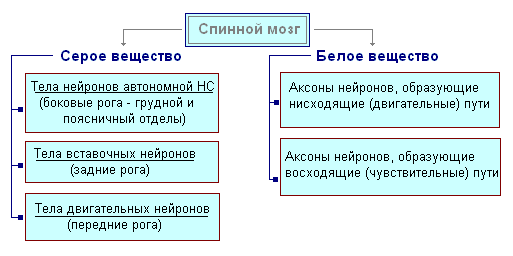
On a transverse section, the gray matter resembles the letter H. It distinguishes between the anterior and posterior horns, as well as the connecting crossbar, in the center of which there is a narrow spinal canal containing cerebrospinal fluid. Lateral horns are isolated in the thoracic region. They contain the bodies of neurons that innervate the internal organs. The white matter of the spinal cord is formed by nerve processes. Short processes connect parts of the spinal cord, and long ones make up the conductor apparatus of bilateral connections with the brain.
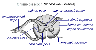
The spinal cord has two thickenings - cervical and lumbar, from which the nerves extend to the upper and lower extremities. There are 31 pairs of spinal nerves that emerge from the spinal cord. Each nerve starts from the spinal cord with two roots - anterior and posterior. back roots - sensitive composed of processes of centripetal neurons. Their bodies are located in the spinal nodes. Front roots - motor- are processes of centrifugal neurons located in the gray matter of the spinal cord. As a result of the fusion of the anterior and posterior roots, a mixed spinal nerve is formed. In the spinal cord centers are concentrated that regulate the simplest reflex acts. The main functions of the spinal cord are reflex activity and conduction of excitation.
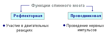
The human spinal cord contains the reflex centers of the muscles of the upper and lower extremities, sweating and urination. The function of conducting excitation is that impulses pass through the spinal cord from the brain to all areas of the body and vice versa. Centrifugal impulses from organs (skin, muscles) are transmitted to the brain along the ascending pathways. Centrifugal impulses are transmitted along descending paths from the brain to the spinal cord, then to the periphery, to the organs. If the pathways are damaged, there is a loss of sensitivity in various parts of the body, a violation of voluntary muscle contractions and the ability to move.
Evolution of the Vertebrate Brain
The formation of the central nervous system in the form of a neural tube first appears in chordates. At lower chordates neural tube persists throughout life higher- vertebrates - in the embryonic stage, the neural plate is laid on the dorsal side, which plunges under the skin and folds into a tube. In the embryonic stage of development, the neural tube forms three swellings in the anterior part - three cerebral vesicles, from which the brain regions develop: the anterior vesicle gives forebrain and diencephalon, middle vesicle turns into midbrain, posterior vesicle forms cerebellum and medulla oblongata. These five parts of the brain are characteristic of all vertebrates.
For lower vertebrates- fish and amphibians - the predominance of the midbrain over the rest of the departments is characteristic. At amphibians the forebrain increases somewhat and a thin layer of nerve cells is formed in the roof of the hemispheres - the primary cerebral fornix, the ancient cortex. At reptiles the forebrain is significantly enlarged due to accumulations of nerve cells. Most of the roof of the hemispheres is occupied by the ancient crust. For the first time in reptiles, the rudiment of a new bark appears. The hemispheres of the forebrain crawl onto other departments, as a result of which a bend is formed in the region of the diencephalon. Since the ancient reptiles, the cerebral hemispheres have become the largest part of the brain.
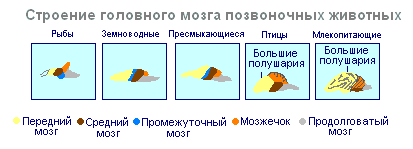
in the structure of the brain birds and reptiles much in common. On the roof of the brain is the primary cortex, the midbrain is well developed. However, in birds, compared with reptiles, the total mass of the brain and the relative size of the forebrain increase. The cerebellum is large and has a folded structure. At mammals the forebrain reaches its greatest size and complexity. Most of the medulla is the new cortex, which serves as the center of higher nervous activity. The intermediate and middle sections of the brain in mammals are small. The growing hemispheres of the forebrain cover them and crush them under them. In some mammals, the brain is smooth, without furrows and convolutions, but in most mammals there are furrows and convolutions in the cerebral cortex. The appearance of furrows and convolutions occurs due to the growth of the brain with a limited size of the skull. Further growth of the cortex leads to the appearance of folding in the form of furrows and convolutions.
Brain
If the spinal cord in all vertebrates is developed more or less equally, then the brain differs significantly in size and complexity of structure in different animals. The forebrain undergoes especially dramatic changes in the course of evolution. In lower vertebrates, the forebrain is poorly developed. In fish, it is represented by the olfactory lobes and nuclei of gray matter in the thickness of the brain. The intensive development of the forebrain is associated with the emergence of animals on land. It differentiates into the diencephalon and into two symmetrical hemispheres called telencephalon. Gray matter on the surface of the forebrain (cortex) first appears in reptiles, developing further in birds and especially in mammals. Indeed, large hemispheres of the forebrain become only in birds and mammals. In the latter, they cover almost all other parts of the brain.
The brain is located in the cranial cavity. It includes the brainstem and telencephalon (cerebral cortex).

brain stem consists of the medulla oblongata, pons, midbrain and diencephalon.
Medulla is a direct continuation of the spinal cord and expanding, passes into the hindbrain. It basically preserves the shape and structure of the spinal cord. In the thickness of the medulla oblongata are accumulations of gray matter - the nuclei of the cranial nerves. The rear axle includes cerebellum and pons. The cerebellum is located above the medulla oblongata and has a complex structure. On the surface of the cerebellar hemispheres, the gray matter forms the cortex, and inside the cerebellum, its nuclei. Like the spinal medulla oblongata, it performs two functions: reflex and conduction. However, the reflexes of the medulla oblongata are more complex. This is expressed in the importance in the regulation of cardiac activity, the state of blood vessels, respiration, sweating. The centers of all these functions are located in the medulla oblongata. Here are the centers of chewing, sucking, swallowing, separation of saliva and gastric juice. Despite its small size (2.5–3 cm), the medulla oblongata is a vital part of the CNS. Damage to it can cause death due to the cessation of breathing and heart activity. The conductive function of the medulla oblongata and the pons is to transmit impulses from the spinal cord to the brain and vice versa.
AT midbrain primary (subcortical) centers of vision and hearing are located, which carry out reflex orientation reactions to light and sound stimuli. These reactions are expressed in various movements of the torso, head and eyes in the direction of stimuli. The midbrain consists of the cerebral peduncles and the quadrigemina. The midbrain regulates and distributes the tone (tension) of the skeletal muscles.
diencephalon consists of two departments - thalamus and hypothalamus, each of which consists of a large number of nuclei of the visual tubercles and the hypothalamic region. Through the visual hillocks centripetal impulses are transmitted to the cerebral cortex from all receptors of the body. Not a single centripetal impulse, no matter where it comes from, can pass to the cortex, bypassing the visual tubercles. Thus, through the diencephalon, all receptors are connected with the cerebral cortex. In the hypothalamic region there are centers that affect metabolism, thermoregulation and endocrine glands.
Cerebellum located behind the medulla oblongata. It is made up of gray and white matter. However, unlike the spinal cord and brainstem, the gray matter - the cortex - is located on the surface of the cerebellum, and the white matter is located inside, under the cortex. The cerebellum coordinates movements, makes them clear and smooth, plays an important role in maintaining the balance of the body in space, and also affects muscle tone. When the cerebellum is damaged, a person experiences a drop in muscle tone, movement disorder and a change in gait, speech slows down, etc. However, after some time, movements and muscle tone are restored due to the fact that intact parts of the central nervous system take over the functions of the cerebellum.
Large hemispheres- the largest and most developed part of the brain. In humans, they form the bulk of the brain and are covered with bark over their entire surface. Gray matter covers the outside of the hemispheres and forms the cerebral cortex. The cortex of the human hemispheres has a thickness of 2 to 4 mm and is composed of 6–8 layers formed by 14–16 billion cells, different in shape, size and functions. Under the bark is white matter. It consists of nerve fibers that connect the cortex with the lower sections of the central nervous system and the individual lobes of the hemispheres among themselves.
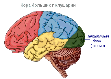
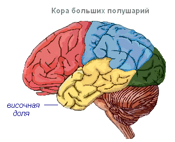
The cerebral cortex has convolutions separated by furrows, which significantly increase its surface. The three deepest furrows divide the hemispheres into lobes. There are four lobes in each hemisphere: frontal, parietal, temporal, occipital. Excitation of different receptors enters the corresponding perceiving areas of the cortex, called zones, and from here are transmitted to a specific organ, prompting it to action. The following zones are distinguished in the cortex. Hearing area located in the temporal lobe, perceives impulses from auditory receptors.

visual area lies in the occipital region. This is where impulses come from the receptors of the eye.
Olfactory zone located on the inner surface of the temporal lobe and is associated with receptors in the nasal cavity.
Sensory-motor zone is located in the frontal and parietal lobes. In this zone are the main centers of movement of the legs, torso, arms, neck, tongue and lips. Here lies the center of speech.

The cerebral hemispheres are the highest division of the central nervous system that controls the functioning of all organs in mammals. The significance of the cerebral hemispheres in humans also lies in the fact that they represent the material basis of mental activity. I.P. Pavlov showed that physiological processes occurring in the cerebral cortex underlie mental activity. Thinking is connected with the activity of the entire cerebral cortex, and not only with the function of its individual areas.
| Department of the brain | Functions | |
| Medulla | Conductor | The connection between the spinal and overlying parts of the brain. |
| reflex | Regulation of the activity of the respiratory, cardiovascular, digestive systems:
|
|
| Pons | Conductor | Connects the hemispheres of the cerebellum to each other and to the cerebral cortex. |
| Cerebellum | Coordinating | Coordination of voluntary movements and maintaining the position of the body in space. Regulation of muscle tone and balance |
| midbrain | Conductor | Orienting reflexes to visual, sound stimuli ( head and body rotations). |
| reflex |
|
|
| diencephalon | thalamus
hypothalamus
|
|
The cerebral cortex
Surface cerebral cortex in humans, it is about 1500 cm 2, which is many times greater than the inner surface of the skull. Such a large surface of the cortex was formed due to the development of a large number of furrows and convolutions, as a result of which most of the cortex (about 70%) is concentrated in the furrows. The largest furrows of the cerebral hemispheres - central, which runs across both hemispheres, and temporal separating the temporal lobe from the rest. The cerebral cortex, despite its small thickness (1.5–3 mm), has a very complex structure. It has six main layers, which differ in the structure, shape and size of neurons and connections. In the cortex there are centers of all sensitive (receptor) systems, representations of all organs and parts of the body. In this regard, centripetal nerve impulses from all internal organs or parts of the body approach the cortex, and it can control their work. Through the cerebral cortex, conditioned reflexes are closed, through which the body constantly, throughout life, very accurately adapts to changing conditions of existence, to the environment.
