❖ The human nervous system is represented by:
■ the brain and spinal cord (together they form central nervous system
);
■ nerves, ganglions and nerve endings (form peripheral part of the nervous system
).
Functions of the human nervous system:
■ unites all parts of the body into a single whole ( integration );
■ regulates and coordinates the work of various organs and systems ( agreement );
■ carries out the connection of the organism with the external environment, its adaptation to environmental conditions and survival in these conditions ( reflection and adaptation );
■ provides (in interaction with the endocrine system) the constancy of the internal environment of the body at a relatively stable level ( correction );
■ determines the consciousness, thinking and speech of a person, his purposeful behavioral, mental and creative activity ( activity ).
❖ Division of the nervous system according to functional characteristics:
■ somatic (innervates the skin and muscles; perceives the effects of the external environment and causes contractions of the skeletal muscles); obeys the will of man;
■ autonomous , or vegetative (regulates metabolic processes, growth and reproduction, the work of the heart and blood vessels, internal organs and endocrine glands).
Spinal cord
Spinal cord located in the spinal canal of the spine, starts from the medulla oblongata (above) and ends at the level of the second lumbar vertebra. It is a white cylindrical cord (cord) with a diameter of about 1 cm and a length of 42-45 cm. The spinal cord has two deep grooves in front and behind, dividing it into the right and left halves.
In the longitudinal direction of the spinal cord, one can distinguish 31 segment , each of which has two front and two back spine formed by axons of neurons; while all segments form a single whole.
Inside spinal cord is located Gray matter , which has (in cross section) the characteristic shape of a flying butterfly, the “wings” of which form front, rear and (in the thoracic region) lateral horns .
Gray matter consists of bodies of intercalary and motor neurons. Along the axis of gray matter along the spinal cord runs a narrow spinal drip , filled cerebrospinal fluid (see below).
On the periphery spinal cord (around gray matter) white matter .
white matter located in the form of 6 columns around the gray matter (two anterior, lateral and posterior).
It is made up of axons assembled in ascending (located in the back and side columns; transmit excitation to the brain) and descending (located in the anterior and lateral columns; transmit excitation from the brain to the working organs) pathways spinal cord.
The spinal cord is protected by rattling sheaths: solid (from the connective tissue that lines the spinal canal) gossamer (in the form of a thin network; contains nerves and vessels) and soft , or vascular (contains many vessels; grows together with the surface of the brain). The space between the arachnoid and soft shells is filled with cerebrospinal fluid, which provides optimal conditions for the vital activity of nerve cells and protects the spinal cord from shocks and concussions.
AT anterior horns segments of the spinal cord (they are located closer to the abdominal surface of the body) are the body motor neurons , from which their axons depart, forming the anterior motor roots , through which excitation is transmitted from the brain to the working organ (these are the longest human cells, their length can reach 1.3 m).
AT posterior horns segments are bodies intercalary neurons ; rear fit them sensitive roots , formed by the axons of sensory neurons that transmit excitation to the spinal cord. The cell bodies of these neurons are located in spinal nodes (ganglia) located outside the spinal cord along the sensory neurons.
In the thoracic region there are lateral horns Where are the bodies of neurons located? sympathetic parts autonomous nervous system.
Outside the spinal canal, the sensory and motor roots extending from the posterior and anterior horns of one "wing" of the segment unite, forming (together with the nerve fibers of the autonomic nervous system) a mixed spinal nerve , which contains both centripetal (sensory) and centrifugal (motor) fibers (see below).
❖ Spinal Cord Functions carried out under the control of the brain.
■ Reflex function:
pass through the gray matter of the spinal cord arcs of unconditioned reflexes
(they do not affect human consciousness), regulating
visceral function, vascular lumen, urination, sexual function, diaphragmatic contraction, defecation, sweating, and managers
skeletal muscles; (examples, knee jerk:
lifting the leg when hitting the tendon attached to the kneecap; limb withdrawal reflex: under the action of a painful stimulus, reflex muscle contraction and limb withdrawal occur; urination reflex: filling the bladder causes excitation of stretch receptors in its wall, which leads to relaxation of the sphincter, contraction of the bladder walls and urination). 
When the spinal cord is ruptured above the arc of the unconditioned reflex, this reflex does not experience the regulatory action of the brain and is perverted (deviates from the norm, i.e. becomes pathological).
■ Conductor function; pathways of the white matter of the spinal cord are conductors of nerve impulses: ascending pathways nerve impulses from the gray matter of the spinal cord go into the brain (nerve impulses coming from sensitive neurons first enter the gray matter of certain segments of the spinal cord, where they undergo preliminary processing), and descending the paths they go from the brain to different segments of the spinal cord and from there along the spinal nerves to the organs.
In humans, the spinal cord controls only simple motor acts; complex movements (walking, writing, labor skills) are carried out with the obligatory participation of the brain.
Paralysis- loss of the ability to voluntary movements of the body's organs, which occurs when the cervical spinal cord is damaged, resulting in a violation of the connection of the brain with the body organs located below the injury site.
spinal shock- this is the disappearance of all reflexes and voluntary movements of the organs of the body, the nerve centers of which lie below the site of injury, arising from injuries of the spine and disruption of communication between the brain and the underlying (in relation to the site of injury) sections of the spinal cord.
Nerves. Propagation of a nerve impulse
Nerves- these are strands of nervous tissue that connect the brain and nerve nodes with other organs and tissues of the body through nerve impulses transmitted through them.
Nerves are formed from several bundles nerve fibers (up to 106 fibers in total) and a small number of thin blood vessels enclosed in a common connective tissue sheath. For each nerve fiber, the nerve impulse propagates in isolation, without passing to other fibers.
■ Most nerves mixed ; they include fibers of both sensory and motor neurons.
nerve fiber- a long (may be more than 1 m long) thin process of a nerve cell ( axon), strongly branching at the very end; serves to transmit nerve impulses.
❖ Classification of nerve fibers depending on the structure: myelinated and unmyelinated .
Myelinated nerve fibers are covered with a myelin sheath. myelin sheath performs the functions of protecting, nourishing and isolating nerve fibers. It has a protein-lipid nature and is a plasmalemma Schwann cell (named after its discoverer T. Schwann, 1810-1882), which repeatedly (up to 100 times) wraps around the axon; while the cytoplasm, all organelles and the shell of the Schwann cell are concentrated on the periphery of the shell above the last turn of the plasmalemma. Between adjacent Schwann cells are open sections of the axon - interceptions of Ranvier . A nerve impulse along such a fiber propagates in jumps from one interception to another at a high speed - up to 120 m / s.
Unmyelinated nerve fibers are covered only by a thin insulating and myelin-free sheath. The speed of propagation of a nerve impulse along an unmyelinated nerve fiber is 0.2–2 m/s.
nerve impulse- This is a wave of excitation that propagates along the nerve fiber in response to irritation of the nerve cell.
■ The speed of propagation of a nerve impulse along a fiber is directly proportional to the square root of the fiber's diameter.
Mechanism of nerve impulse propagation. Simplified, a nerve fiber (axon) can be represented as a long cylindrical tube with a surface membrane separating two aqueous solutions of different chemical composition and concentration. The membrane has numerous valves that close when the electric field increases (i.e., with an increase in its potential difference) and open when it is weakened. In the open state, some of these valves pass Na + ions, other valves pass K + ions, but all of them do not pass large ions of organic molecules.
Each axon is a microscopic power plant, sharing (through chemical reactions) electrical charges. When the axon not excited , inside it there is an excess (compared to the environment surrounding the axon) of potassium cations (K +), as well as negative ions (anions) of a number of organic molecules. Outside the axon there are sodium cations (Na +) and chloride anions (C1 -), which are formed due to the dissociation of NaCl molecules. Anions of organic molecules are concentrated on internal membrane surface, charging it negative , and sodium cations - on its external surface, charging it positively . As a result, an electric field arises between the inner and outer surfaces of the membrane, the potential difference (0.05 V) of which ( resting potential) is large enough to keep the diaphragm valves closed. The resting potential was first described and measured in 1848-1851. German physiologist E.G. Dubois-Reymond in experiments on frog muscles.
When an axon is stimulated, the density of electric charges on its surface decreases, the electric field weakens, and the membrane valves open slightly, allowing the sodium cation Na + into the axon. These cations partially compensate for the negative electric charge of the inner surface of the membrane, as a result of which the direction of the field changes to the opposite at the site of irritation. The process involves neighboring sections of the membrane, which gives rise to the spread of a nerve impulse. At this moment, the valves open, allowing potassium cations K + to pass out, due to which the negative charge inside the axon is gradually restored again, and the potential difference between the inner and outer surfaces of the membrane reaches a value of 0.05 V, characteristic of an unexcited axon. Thus, it is actually not an electric current that propagates along the axon, but a wave of an electrochemical reaction.
■ The shape and speed of propagation of the nerve impulse does not depend on the degree of irritation of the nerve fiber. If it is very strong, there is a whole series of identical impulses; if it is very weak, the impulse does not appear at all. Those. exist some minimum "threshold" degree of stimulation, below which the impulse is not excited.
Impulses entering the neuron along the nerve fiber from any receptor differ only in the number of signals in the series. This means that the neuron only needs to count the number of such signals in one series and, in accordance with the “rules”, how to respond to a given number of consecutive signals, send the necessary command to one or another organ.
spinal nerves
Everyone spinal nerve formed from two roots , extending from the spinal cord: front (efferent) root and rear (afferent) root, which are connected in the intervertebral foramen, forming mixed nerves (contain motor, sensory and sympathetic nerve fibers).
■ A person has 31 pairs of spinal nerves (according to the number of segments of the spinal cord) extending to the right and left of each segment.
Functions of the spinal nerves:
■ they cause sensitivity of the skin of the upper and lower extremities, chest, abdomen;
■ carry out the transmission of nerve impulses that ensure the movement of all parts of the body and limbs;
■ innervate skeletal muscles (diaphragm, intercostal muscles, muscles of the walls of the chest and abdominal cavities), causing their involuntary movements; at the same time, each segment innervates strictly defined areas of the skin and skeletal muscles.
Voluntary movements are carried out under the control of the cerebral cortex.
❖ Innervation by segments of the spinal cord:
■ segments of the cervical and upper thoracic parts of the spinal cord innervate the organs of the chest cavity, heart, lungs, muscles of the head and upper limbs;
■ the remaining segments of the thoracic and lumbar parts of the spinal cord innervate the organs of the upper and middle parts of the abdominal cavity and the muscles of the body;
■ The lower lumbar and sacral segments of the spinal cord innervate the organs of the lower part of the abdominal cavity and the muscles of the lower extremities.
cerebrospinal fluid
cerebrospinal fluid- a transparent, almost colorless liquid containing 89% water. Changes 5 times a day.
❖ Functions of cerebrospinal fluid:
■ creates a mechanical protective "cushion" for the brain;
■ is the internal environment from which the nerve cells of the brain receive nutrients;
■ participates in the removal of exchange products;
■ participates in the maintenance of intracranial pressure.
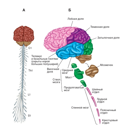
Brain. General characteristics of the structure
Brain located in the cranial cavity and covered with three meninges, equipped with vessels; its mass in an adult is 1100-1700 g.
❖Structure: the brain is made up of 5 departments:
■ medulla oblongata,
■ hindbrain,
■ midbrain,
■ diencephalon,
■ forebrain. 
brain stem - it is a system formed by the medulla oblongata, hindbrain pons, midbrain and diencephalon
In some textbooks and manuals, not only the pons of the hindbrain, but the entire hindbrain, including both the pons varolii and the cerebellum, are referred to the trunk of the brain bridge.
In the brainstem are the nuclei of the cranial nerves that connect the brain with the sense organs, muscles and some glands; gray the substance in it is inside in the form of nuclei, white - outside . White matter consists of processes of neurons that connect parts of the brain to each other.
Bark the cerebral hemispheres and the cerebellum is formed by gray matter, consisting of the bodies of neurons.
Inside the brain are communicating cavities ( cerebral ventricles ), which are a continuation of the central canal of the spinal cord and filled cerebrospinal fluid: I and II lateral ventricles - in the hemispheres of the forebrain, III - in the diencephalon, IV - in the medulla oblongata.
The channel connecting the IV and III ventricles and passing through the midbrain is called aqueduct of the brain.
12 pairs depart from the nuclei of the brain cranial nerves , innervating the sense organs, tissues of the head, neck, organs of the chest and abdominal cavities.
The brain (like the spinal cord) is covered with three shells: solid (from dense connective tissue; performs a protective function), gossamer (contains nerves and vessels) and vascular (contains many vessels). The space between the arachnoid and choroid is filled cerebral fluid .
The existence, location and function of the various centers of the brain are determined by stimulation various structures of the brain electric shock .
Medulla
Medulla is a direct continuation of the spinal cord (after it passes through the foramen magnum) and has a structure similar to it; at the top it borders on the bridge; it contains the fourth ventricle. White matter is located mainly on the outside and forms 2 protrusions - pyramids , the gray matter is located inside the white matter, forming in it numerous nuclei .
■ The nuclei of the medulla oblongata control many vital functions; that's why they are called centers .
❖ Functions of the medulla oblongata:
■ conductive: sensory and motor pathways pass through it, along which impulses are transmitted from the spinal cord to the overlying parts of the brain and back;
■ reflex(carried out together with the pons varolii): in centers the medulla oblongata closes the arcs of many important unconditioned reflexes: respiration and circulation , as well as sucking, salivation, swallowing, gastric secretion (responsible for digestive reflexes ), coughing, sneezing, vomiting, blinking (responsible for defensive reflexes ), etc. Damage to the medulla oblongata leads to cardiac and respiratory arrest and instant death.
Hind brain
Hind brain consists of two departments - pons and cerebellum .
Bridge (Varolian bridge) located between the medulla oblongata and midbrain; Nerve pathways pass through it, connecting the forebrain and midbrain with the medulla oblongata and spinal cord. The facial and auditory cranial nerves depart from the bridge.
❖ Functions of the hindbrain: together with the medulla oblongata, the bridge performs conductive and reflex functions as well governs digestion, respiration, cardiac activity, movement of the eyeballs, contraction of facial muscles that provide facial expressions, etc.
Cerebellum located above the medulla oblongata and consists of two small lateral hemispheres , the middle (most ancient, stem) part, connecting the hemispheres and called cerebellar worm , and three pairs of legs connecting the cerebellum with the midbrain, pons varolii and medulla oblongata.
The cerebellum is covered bark from the gray matter, under which is the white matter; the vermis and cerebellar peduncles also consist of white matter. Within the white matter of the cerebellum are nuclei made up of gray matter. The cerebellar cortex has numerous elevations (gyrus) and depressions (sulci). Most cortical neurons are inhibitory.
❖ Functions of the cerebellum:
■ the cerebellum receives information from the muscles, tendons, joints and motor centers of the brain;
■ it ensures the maintenance of muscle tone and body posture,
■ coordinates body movements (makes them accurate and coordinated);
■ manages balance.
With the destruction of the cerebellar vermis, a person cannot walk and stand, with damage to the hemispheres of the cerebellum, speech and writing are disturbed, severe trembling of the limbs appears, movements of the arms and legs become sharp.
Reticular Formation
Reticular (mesh) formation- This is a dense network formed by a cluster of neurons of different sizes and shapes, with well-developed processes that run in different directions and many synaptic contacts.
■ The reticular formation is located in the middle part of the medulla oblongata, in the pons and midbrain.
❖ Functions of the reticular formation:
■ its neurons sort (pass, delay or supply additional energy) incoming nerve impulses;
■ it regulates the excitability of all parts of the nervous system located above it ( ascending influences ) and below ( downward influences ), and is a center that stimulates the centers of the cerebral cortex;
■ the state of wakefulness and sleep is associated with its activity;
■ it ensures the formation of sustainable attention, emotions, thinking and consciousness;
■ with its participation, the regulation of digestion, respiration, heart activity, etc. is carried out.
midbrain
midbrain- the smallest part of the brain located above the bridge between the diencephalon and the cerebellum. Introduced quadrigemina (2 upper and 2 lower tubercles) and legs of the brain . There is a canal in its center water pipes ), connecting the III and IV ventricles and filled with cerebrospinal fluid.
❖ Midbrain functions:
■conductive: in its legs there are ascending nerve pathways to the cerebral cortex and cerebellum and descending nerve pathways along which impulses go from the cerebral hemispheres and cerebellum to the medulla oblongata and spinal cord;
■ reflex: it is associated with reflexes of the body posture, its rectilinear movement, rotation, lifting, descent and landing, arising with the participation of the sensory balance system and providing coordination of movement in space;
■ in the quadrigemina there are subcortical centers of visual and auditory reflexes that provide orientation towards sound and light. The neurons of the superior colliculus of the quadrigemina receive impulses from the eyes and muscles of the head and respond to objects moving rapidly in the field of view; neurons of the inferior colliculus respond to strong, sharp sounds, putting the auditory system on high alert;
■ it regulates muscle tone , provides fine finger movements, chewing.
diencephalon
❖ diencephalon- this is the final section of the brain stem; it is located under the cerebral hemispheres of the forebrain above the midbrain. It contains centers that process nerve impulses entering the cerebral hemispheres, as well as centers that control the activity of internal organs.
The structure of the diencephalon: it consists of the central part - thalamus (visual tubercles), hypothalamus (subtubercular region) and cranked bodies ; it also contains the third ventricle of the brain. Located at the base of the hypothalamus pituitary.
■ thalamus- this is a kind of "control room", through which all information about external environment and state of the body. The thalamus controls the rhythmic activity of the cerebral hemispheres, is the subcortical center for analysis of all types sensations , except for olfactory; it houses the centers that regulate sleep and wakefulness, emotional reactions(feelings of aggression, pleasure and fear) and mental activity person. AT ventral nuclei thalamus is formed sensation pain and maybe feeling time .
If the thalamus is damaged, the nature of sensations can change: for example, even slight touches on the skin, sound or light can cause severe attacks of pain in a person; on the contrary, sensitivity may decrease so much that a person will not respond to any irritation.
■ Hypothalamus- the highest center of vegetative regulation. He perceives changes in the internal environment body and regulates metabolism, body temperature, blood pressure, homeostasis, endocrine glands. It has centers hunger, satiety, thirst, regulation body temperature etc. It releases biologically active substances ( neurohormones ) and substances necessary for the synthesis of neurohormones pituitary gland , carrying out neurohumoral regulation the vital activity of the organism. The anterior nuclei of the hypothalamus are the center of parasympathetic autonomic regulation, the posterior nuclei are sympathetic.
■ Pituitary- lower appendage of the hypothalamus; is an endocrine gland (for details, see "").
Forebrain. The cerebral cortex
❖ forebrain represented by two large hemispheres and corpus callosum connecting the hemispheres. The large hemispheres control the work of all organ systems and provide the relationship of the body with the external environment. The corpus callosum plays an important role in the processing of information in the learning process.
big hemispheres two - solder and left ; they cover the midbrain and diencephalon. In an adult, the cerebral hemispheres account for up to 80% of the mass of the brain.
On the surface of each hemisphere there are many furrows (recesses) and convolutions (folds).
Main furrows; central, lateral and parietal-occipital. Furrows divide each hemisphere into 4 shares (see below); which, in turn, are divided by furrows into a series convolutions .
Inside the cerebral hemispheres are the 1st and 2nd ventricles of the brain.
The major hemispheres are covered gray matter - bark , consisting of several layers of neurons that differ from each other in shape, size and function. In total, there are 12-18 billion bodies of neurons in the cerebral cortex. The thickness of the bark is 1.5-4.5 mm, the area is 1.7-2.5 thousand cm2. Furrows and convolutions significantly increase the surface area and volume of the cortex (2/3 of the cortical area is hidden in the furrows).
The right and left hemispheres are functionally different from each other ( functional asymmetry of the hemispheres ). The presence of functional asymmetry of the hemispheres was established in experiments on people with a "split brain".
■ Operation " brain splitting a" consists in the surgical cutting (for medical reasons) of all direct connections between the hemispheres, as a result of which they begin to function independently of each other.
At right-handers the leading (dominant) hemisphere is left , and at left-handed - right .
■ Right hemisphere responsible for creative thinking , forms the basis creativity , acceptance non-standard solutions . Damage to the visual zone of the right hemisphere leads to impaired face recognition.
■ Left hemisphere provides logical reasoning and abstract thinking (the ability to operate with mathematical formulas, etc.), it contains centers oral and written speeches , formation decisions . Damage to the visual zone of the left hemisphere leads to impaired recognition of letters and numbers.
Despite its functional asymmetry, the brain functions as whole , providing consciousness, memory, thinking, adequate behavior, various types of conscious human activity.
❖ Functions of the cortex cerebral hemispheres:
■ carries out higher nervous activity (consciousness, thinking, speech, memory, imagination, the ability to write, read, count);
■ provides the relationship of the body with the external environment, is the central department of all analyzers; various sensations are formed in its zones (the zones of hearing and taste are located in the temporal lobe; vision - in the occipital; speech - in the parietal and temporal; skin-muscle sense - in the parietal; movement - in the frontal);
■ provides mental activity;
■ arcs of conditioned reflexes are closed in it (ie it is an organ for acquiring and accumulating life experience).
❖Lobes of the bark- subdivision of the surface of the cortex according to the anatomical principle: in each hemisphere, the frontal, temporal, parietal and occipital lobes are distinguished. 
❖ Cortex zone- a section of the cerebral cortex, characterized by the uniformity of the structure and functions performed.
❖ Types of cortical zones: sensory (or projection), associative, motor.
Sensory or projection zones- these are the highest centers of various types of sensitivity; when they are irritated, the simplest sensations arise, and when damaged, a violation of sensory functions occurs (blindness, deafness, etc.). These zones are located in the areas of the cortex, where the ascending pathways end, along which nerve impulses from the receptors of the sense organs (visual zone, auditory zone, etc.) are conducted.
■ visual area located in the occipital region of the cortex;
■olfactory, gustatory and auditory areas - in the temporal region and next to it;
■ skin and muscle sensation zones - in the posterior central gyrus.
Association zones- areas of the cortex responsible for generalized information processing; processes that ensure the mental functions of a person take place in them - thinking, speech, emotions, etc.
In associative zones, excitation occurs when impulses arrive not only in these, but also in sensory zones, and not only from one, but also simultaneously from several sense organs (for example, excitation in the visual zone can appear in response not only to visual, but also to auditory stimuli).
■ Frontal associative areas of the cortex provide the development of sensory information and form the goal and program of action, consisting of commands sent to the executive organs. From these organs, the frontal associative zones receive feedback about the implementation of actions and their direct consequences. In the frontal associative zones, this information is analyzed, it is determined whether the goal has been achieved, and if it is not achieved, the commands to the organs are corrected.
■ The development of the frontal lobes of the cortex to a large extent determined the high level of human mental abilities in comparison with primates.
Motor (motor) zones- areas of the cortex, irritation of which causes muscle contraction. These zones control voluntary movements; they originate descending conducting paths along which nerve impulses go to the intercalary and executive neurons.
■ The motor function of various parts of the body is represented in the anterior central gyrus. The largest space is occupied by the motor zones of the hands, fingers and muscles of the face, the smallest - by the zones of the muscles of the body.
Electroencephalogram
Electroencephalogram (EEG)- this is a graphical record of the total electrical activity of the cerebral cortex - nerve impulses generated by a combination of its (cortex) neurons.
■ In the human EEG, waves of electrical activity of different frequencies are observed - from 0.5 to 30 oscillations per second.
Basic rhythms of electrical activity cerebral cortex: alpha rhythm, beta rhythm, delta rhythm and theta rhythm.
alpha rhythm- oscillations with a frequency of 8-13 hertz; this rhythm prevails over others during sleep.
beta rhythm has an oscillation frequency of more than 13 hertz; it is characteristic of active wakefulness.
Theta rhythm- oscillations with a frequency of 4-8 hertz.
delta rhythm has a frequency of 0.5-3.5 hertz.
■ Theta and delta rhythms are observed during very deep sleep or anesthesia .
cranial nerves
cranial nerves a person has 12 pairs; they depart from different parts of the brain and are divided by function into sensory, motor and mixed.
❖ Sensitive nerves-1, II, VIII couples:
■ I couple — olfactory nerves that depart from the forebrain and innervate the olfactory region of the nasal cavity;
■ And couple — visual nerves that depart from the diencephalon and innervate the retina of the eye;
■ VIII pair - auditory (or vestibulocochlear e) nerves; depart from the bridge, innervate the membranous labyrinth and the Cor-ti's organ of the inner ear.
❖ Motor nerves- III, IV, VI, X, XII pairs:
■ III pair — oculomotor nerves arising from the midbrain;
■ IV pair — blocky nerves also arise from the midbrain;
■ VI - diverting nerves that depart from the bridge (III, IV and VI pairs of nerves innervate the muscles of the eyeball and eyelids);
■ XI - additional nerves, depart from the medulla oblongata;
■XII— sublingual nerves also depart from the medulla oblongata (XI and XII pairs of nerves innervate the muscles of the pharynx, tongue, middle ear, parotid salivary gland).
❖ mixed nerves-V, VII, IX, X pairs:
■ V pair — trigeminal nerves that depart from the bridge, innervate the scalp, eye membranes, masticatory muscles, etc .;
■ VII pair - facial nerves also depart from the bridge, innervate the facial muscles, the lacrimal gland, etc .;
■ IX couple — glossopharyngeal nerves that depart from the diencephalon, innervate the muscles of the pharynx, middle ear, parotid salivary gland;
■ X pair — wandering nerves also depart from the diencephalon, innervate the muscles of the soft palate and larynx, the organs of the chest (trachea, bronchi, heart, slowing down its work) and abdominal cavities (stomach, liver, pancreas).
Features of the autonomic nervous system
Unlike the somatic nervous system, the nerve fibers of which are thick, covered with a myelin sheath and characterized by a high speed of propagation of nerve impulses, autonomic nerve fibers are usually thin, do not have a myelin sheath and are characterized by a low speed of propagation of nerve impulses (see table). 
❖ Functions of the autonomic nervous system:
■ maintaining the constancy of the internal environment of the body through the neuroregulation of tissue metabolism ("start", correction or suspension of certain metabolic processes) and the work of internal organs, the heart and blood vessels;
■ adaptation of the activities of these organs to the changed environmental conditions and the needs of the body.
The autonomic nervous system is made up of sympathetic and parasympathetic parts , which have the opposite effect on the physiological functions of organs.
sympathetic part The autonomic nervous system creates conditions for intensive activity of the body, especially in extreme conditions, when it is necessary to demonstrate all the capabilities of the body.
parasympathetic part(the "retreat" system) of the autonomic nervous system reduces the level of activity, which contributes to the restoration of resources spent by the body.
■ Both parts (sections) of the autonomic nervous system are subordinate to higher nerve centers located in hypothalamus , and complement each other.
■ The hypothalamus coordinates the work of the autonomic nervous system with the activity of the endocrine and somatic systems.
■ Examples of the influence of the sympathetic and parasympathetic parts of the ANS on the organs are given in the table on p. 520.
The effective performance of the functions of both parts of the autonomic nervous system is ensured double innervation internal organs and heart.
double innervation internal organs and the heart means that nerve fibers from both the sympathetic and parasympathetic parts of the autonomic nervous system approach each of these organs. 
Neurons of the autonomic nervous system synthesize various mediators (acetylcholine, norepinephrine, serotonin, etc.) involved in the transmission of nerve impulses.
main feature autonomic nervous system - bineuronality of the efferent pathway
. This means that in the autonomic nervous system efferent
, or centrifugal
(i.e. coming from the head and spinal brain to organs
), nerve impulses sequentially pass through the bodies of two neurons. The two-neuronality of the efferent pathway makes it possible to distinguish in the sympathetic and parasympathetic parts of the autonomic nervous system central and peripheral parts
.
central part (nerve centers ) autonomic nervous system located in the central nervous system (in the lateral horns of the gray matter of the spinal cord, as well as in the medulla oblongata and midbrain) and contains the first motor neurons of the reflex arc . The autonomic nerve fibers going from these centers to the working organs switch in the autonomic ganglia of the peripheral part of the autonomic nervous system.
peripheral part The autonomic nervous system is located outside the central nervous system and consists of ganglion (nerve ganglions) formed by the bodies second motor neurons of the reflex arc as well as nerves and nerve plexuses.
■ At sympathetic department, these ganglia form a pair sympathetic chains (trunks) located near the spine on both sides of it, in the parasympathetic department they lie near or inside the innervated organs.
■ Postganglionic parasympathetic fibers approach the eye muscles, larynx, trachea, lungs, heart, lacrimal and salivary glands, muscles and glands of the digestive tract, excretory and genital organs. 
Causes of disruption of the nervous system
❖ Overwork of the nervous system weakens its regulatory function and can provoke the occurrence of a number of mental, cardiovascular, gastrointestinal, skin and other diseases.
❖ hereditary diseases can lead to changes in the activity of some enzymes. As a result, toxic substances accumulate in the body, the impact of which leads to impaired brain development and mental retardation.
Negative environmental factors:
■bacterial infections lead to the accumulation of toxins in the blood, poisoning the nervous tissue (meningitis, tetanus);
■ viral infections can affect the spinal cord (poliomyelitis) or the brain (encephalitis, rabies);
■ alcohol and its metabolic products excite various nerve cells (inhibitory or excitatory neurons), disorganizing the work of the nervous system; the systematic use of alcohol causes chronic depression of the nervous system, changes in skin sensitivity, muscle pain, weakening and even disappearance of many reflexes; irreversible changes occur in the central nervous system, forming personality changes and leading to the development of severe mental illness and dementia;
■ influence nicotine and drugs much like the effect of alcohol;
■heavy metal salts bind to enzymes, disrupting their work, which leads to disruption of the nervous system;
■ when bites of poisonous animals biologically active substances (poisons) that disrupt the functioning of neuronal membranes enter the bloodstream;
■ when head injuries, bleeding and severe pain possible loss of consciousness, which is preceded by: darkening in the eyes, tinnitus, pallor, lowering the temperature, profuse sweating, weak pulse, shallow breathing.
Violation of cerebral circulation. The narrowing of the lumen of the brain vessels leads to disruption of the normal functioning of the brain and, as a result, to diseases of various organs. Injuries and high blood pressure can cause rupture of cerebral vessels, which usually leads to paralysis, higher nervous activity disorders, or death.
Clamping of the nerve trunks of the brain causes severe pain. Infringement of the roots of the spinal cord by spasmodic back muscles or as a result of inflammation causes paroxysmal pain (typical for sciatica ), sensory disturbance ( numbness ) and etc.
❖ When metabolic disorders in the brain mental illness occurs
■neurosis - emotional, motor and behavioral disorders, accompanied by deviations from the autonomic nervous system and the work of internal organs (example: fear of the dark in children);
■ affective insanity - a more serious illness in which periods of extreme excitement alternate with apathy (paranoia, megalomania or persecution);
■ schizophrenia - splitting of consciousness;
■ hallucinations (may also occur with poisoning, high fever, acute alcoholic psychosis).
The human nervous system is similar in structure to the nervous system of higher mammals, but differs in a significant development of the brain. The main function of the nervous system is to control the vital activity of the whole organism.
Neuron
All organs of the nervous system are built from nerve cells called neurons. A neuron is capable of receiving and transmitting information in the form of a nerve impulse.

Rice. 1. Structure of a neuron.
The body of a neuron has processes by which it communicates with other cells. The short processes are called dendrites, the long ones are called axons.
The structure of the human nervous system
The main organ of the nervous system is the brain. It is connected to the spinal cord, which looks like a cord about 45 cm long. Together, the spinal cord and brain make up the central nervous system (CNS).

Rice. 2. Scheme of the structure of the nervous system.
Nerves leaving the CNS make up the peripheral part of the nervous system. It consists of nerves and nerve nodes.
TOP 4 articleswho read along with this
Nerves are formed from axons, the length of which can exceed 1 m.
Nerve endings contact each organ and transmit information about their condition to the central nervous system.
There is also a functional division of the nervous system into somatic and autonomic (autonomous).
The part of the nervous system that innervates the striated muscles is called the somatic. Her work is connected with the conscious efforts of a person.
The autonomic nervous system (ANS) regulates:
- circulation;
- digestion;
- selection;
- breath;
- metabolism;
- smooth muscle work.
Thanks to the work of the autonomic nervous system, there are many processes of normal life that we do not consciously regulate and usually do not notice.
The significance of the functional division of the nervous system is in ensuring the normal, independent of our consciousness, functioning of the finely tuned mechanisms of the work of internal organs.
The highest organ of the ANS is the hypothalamus, located in the intermediate part of the brain.
The ANS is divided into 2 subsystems:
- sympathetic;
- parasympathetic.
Sympathetic nerves activate the organs and control them in situations that require action and increased attention.
Parasympathetic slow down the work of the organs and turn on during rest and relaxation.
For example, sympathetic nerves dilate the pupil, stimulate salivation. Parasympathetic, on the contrary, narrow the pupil, slow down salivation.
Reflex
This is the response of the body to irritation from the external or internal environment.
The main form of activity of the nervous system is a reflex (from the English reflection - reflection).
An example of a reflex is pulling the hand away from a hot object. The nerve ending perceives high temperature and transmits a signal about it to the central nervous system. In the central nervous system, a response impulse arises, going to the muscles of the arm.

Rice. 3. Scheme of the reflex arc.
Sequence: sensory nerve - CNS - motor nerve is called the reflex arc.
Brain
The brain is characterized by a strong development of the cerebral cortex, in which the centers of higher nervous activity are located.
The features of the human brain sharply separated it from the animal world and allowed it to create a rich material and spiritual culture.
What have we learned?
The structure and functions of the human nervous system are similar to those of mammals, but differ in the development of the cerebral cortex with the centers of consciousness, thinking, memory, and speech. The autonomic nervous system controls the body without the participation of consciousness. The somatic nervous system controls the movement of the body. The principle of activity of the nervous system is reflex.
Topic quiz
Report Evaluation
Average rating: 4.4. Total ratings received: 380.
The nervous system controls the activity of all systems and organs and ensures the connection of the body with the external environment.
The structure of the nervous system
The structural unit of the nervous system is the neuron - a nerve cell with processes. In general, the structure of the nervous system is a collection of neurons that are constantly in contact with each other using special mechanisms - synapses. The following types of neurons differ in function and structure:
- Sensitive or receptor;
- Effector - motor neurons that send an impulse to the executive organs (effectors);
- Closing or plug-in (conductor).
Conventionally, the structure of the nervous system can be divided into two large sections - somatic (or animal) and vegetative (or autonomous). The somatic system is primarily responsible for the connection of the body with the external environment, providing movement, sensitivity and contraction of skeletal muscles. The vegetative system affects the growth processes (respiration, metabolism, excretion, etc.). Both systems have a very close relationship, only the autonomic nervous system is more independent and does not depend on the will of a person. That is why it is also called autonomous. The autonomous system is divided into sympathetic and parasympathetic.
The entire nervous system consists of the central and peripheral. The central part includes the spinal cord and brain, and the peripheral system represents the outgoing nerve fibers from the brain and spinal cord. If you look at the brain in section, you can see that it consists of white and gray matter.
Gray matter is an accumulation of nerve cells (with the initial sections of processes extending from their bodies). Separate groups of gray matter are also called nuclei.
White matter consists of nerve fibers covered with myelin sheath (processes of nerve cells from which gray matter is formed). In the spinal cord and brain, nerve fibers form pathways.
Peripheral nerves are divided into motor, sensory and mixed, depending on what fibers they consist of (motor or sensory). The bodies of neurons, whose processes are made up of sensory nerves, are located in ganglions outside the brain. The bodies of motor neurons are located in the motor nuclei of the brain and the anterior horns of the spinal cord.
Functions of the nervous system
The nervous system has different effects on the organs. The three main functions of the nervous system are:
- Starting, causing or stopping the function of an organ (secretion of the gland, muscle contraction, etc.);
- Vasomotor, which allows you to change the width of the lumen of the vessels, thereby regulating the flow of blood to the organ;
- Trophic, lowering or increasing metabolism, and, consequently, the consumption of oxygen and nutrients. This allows you to constantly coordinate the functional state of the body and its need for oxygen and nutrients. When impulses are sent along the motor fibers to the working skeletal muscle, causing its contraction, then impulses are simultaneously received that increase metabolism and dilate blood vessels, which makes it possible to provide energy for performing muscle work.
Diseases of the nervous system
Together with the endocrine glands, the nervous system plays a crucial role in the functioning of the body. It is responsible for the coordinated work of all systems and organs of the human body and unites the spinal cord, brain and peripheral system. Motor activity and sensitivity of the body is supported by nerve endings. And thanks to the autonomic system, the cardiovascular system and other organs are inverted.
Therefore, a violation of the functions of the nervous system affects the work of all systems and organs.
All diseases of the nervous system can be divided into infectious, hereditary, vascular, traumatic and chronically progressive.
Hereditary diseases are genomic and chromosomal. The most famous and common chromosomal disease is Down's disease. This disease is characterized by the following symptoms: a violation of the musculoskeletal system, the endocrine system, lack of mental abilities.
Traumatic lesions of the nervous system occur due to bruises and injuries, or when squeezing the brain or spinal cord. Such diseases are usually accompanied by vomiting, nausea, memory loss, disorders of consciousness, loss of sensitivity.
Vascular diseases mainly develop against the background of atherosclerosis or hypertension. This category includes chronic cerebrovascular insufficiency, cerebrovascular accident. Characterized by the following symptoms: attacks of vomiting and nausea, headache, impaired motor activity, decreased sensitivity.
Chronically progressive diseases, as a rule, develop as a result of metabolic disorders, exposure to infection, intoxication of the body, or due to abnormalities in the structure of the nervous system. Such diseases include sclerosis, myasthenia, etc. These diseases usually progress gradually, reducing the efficiency of some systems and organs.
Causes of diseases of the nervous system:
The placental route of transmission of diseases of the nervous system during pregnancy (cytomegalovirus, rubella), as well as through the peripheral system (poliomyelitis, rabies, herpes, meningoencephalitis) is also possible.
In addition, the nervous system is negatively affected by endocrine, heart, kidney diseases, malnutrition, chemicals and drugs, heavy metals.
The entire nervous system is divided into central and peripheral. The central nervous system includes the brain and spinal cord. From them, nerve fibers diverge throughout the body - the peripheral nervous system. It connects the brain with the sense organs and with the executive organs - the muscles and glands.
All living organisms have the ability to respond to physical and chemical changes in the environment.
Stimuli of the external environment (light, sound, smell, touch, etc.) are converted by special sensitive cells (receptors) into nerve impulses - a series of electrical and chemical changes in the nerve fiber. Nerve impulses are transmitted along sensitive (afferent) nerve fibers to the spinal cord and brain. Here, the corresponding command impulses are generated, which are transmitted along the motor (efferent) nerve fibers to the executive organs (muscles, glands). These executive organs are called effectors.
The main function of the nervous system is the integration of external influences with the corresponding adaptive reaction of the body.
The structural unit of the nervous system is the nerve cell, the neuron. It consists of a cell body, a nucleus, branched processes - dendrites - along them nerve impulses go to the cell body - and one long process - an axon - along it a nerve impulse passes from the cell body to other cells or effectors.
The processes of two adjacent neurons are connected by a special formation - a synapse. It plays an essential role in filtering nerve impulses: it passes some impulses and delays others. Neurons are connected to each other and carry out joint activities.
The central nervous system consists of the brain and spinal cord. The brain is divided into the brainstem and the forebrain. The brain stem consists of the medulla oblongata and midbrain. The forebrain is divided into intermediate and final.
All parts of the brain have their own functions.
Thus, the diencephalon consists of the hypothalamus - the center of emotions and vital needs (hunger, thirst, libido), the limbic system (in charge of emotional-impulsive behavior) and the thalamus (which performs filtering and primary processing of sensory information).
In humans, the cerebral cortex is especially developed - the organ of higher mental functions. It has a thickness of 3 mm, and its total area is on average 0.25 sq.m.
The bark is made up of six layers. The cells of the cerebral cortex are interconnected.
There are about 15 billion of them.
Different cortical neurons have their own specific function. One group of neurons performs the function of analysis (crushing, dismemberment of a nerve impulse), the other group performs synthesis, combines impulses coming from various sensory organs and parts of the brain (associative neurons). There is a system of neurons that keeps traces of previous influences and compares new influences with existing traces.
According to the features of the microscopic structure, the entire cerebral cortex is divided into several dozen structural units - fields, and according to the location of its parts - into four lobes: occipital, temporal, parietal and frontal.
The human cerebral cortex is a holistically working organ, although some of its parts (areas) are functionally specialized (for example, the occipital region of the cortex performs complex visual functions, the frontal-temporal - speech, temporal - auditory). The largest part of the motor zone of the human cerebral cortex is associated with the regulation of the movement of the labor organ (hand) and speech organs.
All parts of the cerebral cortex are interconnected; they are also connected to the underlying parts of the brain, which carry out the most important vital functions. Subcortical formations, regulating innate unconditional reflex activity, are the area of those processes that are subjectively felt in the form of emotions (they, according to I.P. Pavlov, are “a source of strength for cortical cells”).
The human brain contains all the structures that arose at various stages of the evolution of living organisms. They contain the "experience" accumulated in the process of the entire evolutionary development. This testifies to the common origin of man and animals.
As the organization of animals at various stages of evolution becomes more complex, the importance of the cerebral cortex grows more and more.
If, for example, the cerebral cortex of a frog is removed (it has an insignificant share in the total volume of its brain), then the frog almost does not change its behavior. Deprived of the cerebral cortex, the dove flies, maintains balance, but already loses a number of vital functions. A dog with a removed cerebral cortex becomes completely unadapted to the environment.
The main mechanism of nervous activity is the reflex. Reflex
The reaction of the body to external or internal influences through the central nervous system.
The term "reflex", as already noted, was introduced into physiology by the French scientist Rene Descartes in the 17th century. But to explain mental activity, it was used only in 1863 by the founder of Russian materialistic physiology, M.I. Sechenov. Developing the teachings of I.M. Sechenov, I.P. Pavlov experimentally investigated the features of the functioning of the reflex.
All reflexes are divided into two groups: conditioned and unconditioned.
Unconditioned reflexes are innate reactions of the body to vital stimuli (food, danger, etc.). They do not require any conditions for their development (for example, the blink reflex, salivation at the sight of food).
Unconditioned reflexes are a natural reserve of ready-made, stereotyped reactions of the body. They arose as a result of a long evolutionary development of this species of animals. Unconditioned reflexes are the same in all individuals of the same species; it is the physiological mechanism of instincts. But the behavior of higher animals and humans is characterized not only by innate, i.e. unconditional reactions, but also such reactions that are acquired by a given organism in the course of its individual life activity, i.e. conditioned reflexes.
Conditioned reflexes are a physiological mechanism for adapting the body to changing environmental conditions.
Conditioned reflexes are such reactions of the body that are not innate, but are developed in various lifetime conditions.
They arise under the condition of constant precedence of various phenomena to those that are vital for the animal. If the connection between these phenomena disappears, then the conditioned reflex fades (for example, the growl of a tiger in a zoo, without being accompanied by its attack, ceases to frighten other animals).
The brain does not go on about only current influences. He plans, anticipates the future, carries out an anticipatory reflection of the future. This is the main feature of his work. The action must achieve a certain future result - the goal. Without preliminary modeling by the brain of this result, regulation of behavior is impossible.
The modern science of the brain - neurophysiology - is based on the concept of the functional combination of brain mechanisms for the implementation of behavioral acts. This concept was put forward and fruitfully developed by the student of I.P. Pavlov, Academician P.K. Anokhin in his theory of functional systems.
Functional system P.K. Anokhin calls the unity of central and peripheral neurophysiological mechanisms, which together ensure the effectiveness of a behavioral act.
The initial stage of formation of any behavioral act was named by P.K.
In the process of afferent synthesis, various information is processed, coming from the external and internal world, on the basis of the currently dominant motivation (need). From the numerous formations of the brain, everything that was connected in the past with the satisfaction of this need is extracted.
Establishing that a given need can be satisfied by a certain action, choosing this action is called making a decision.
The neurophysiological decision-making mechanism is called by P.K. Anokhin the acceptor of the results of action. The acceptor (“assertare”-permissive) of the results of an action is a neurophysiological mechanism for predicting the results of a future action. Based on a comparison of previously obtained results, an action program is created. And only after that the action itself takes place. The course of action, the effectiveness of its stages, the correspondence of these results to the formed program of action is constantly monitored by receiving signals about the achievement of the goal. This mechanism of constant receipt of information about the results of the performed action is called by P.K. Anokhin the reverse afferentation.
The nervous system plays an exceptional integrating role in the life of the organism, as it unites (integrates) it into a single whole and "fits" (integrates) it into the environment. It ensures the coordinated work of individual parts of the body ( coordination), maintaining an equilibrium state in the body ( homeostasis) and adaptation of the organism to changes in the external and / or internal environment ( adaptive state and/or adaptive behavior).
The most important thing that the nervous system does
The nervous system provides the relationship and interaction between the body and the external environment. And for this she does not need so many processes.
Basic processes in the nervous system
1. transduction . The transformation of a stimulus external to the nervous system itself into a nervous excitation with which it can operate.
2. Transformation . Alteration, transformation of the incoming excitation stream into an outgoing stream with different characteristics.
3. Distribution . The distribution of excitation and its direction along different paths, to different addresses.
4. Modeling. Construction of a neural model of stimulation and/or stimulus that replaces the stimulus itself. The nervous system can work with this model, it can store it, modify it and use it instead of a real stimulus. Sensory image is one of the variants of neural models of stimulation.
5. Modulation . The nervous system under the influence of irritation changes itself and / or its activity.
Types of modulation
1. Activation (excitation). An increase in the activity of the nervous structure, an increase in its excitation and / or excitability. dominant state.
2. Oppression (inhibition, inhibition). Decreased activity of the nervous structure, inhibition.
3. Plastic restructuring of the nervous structure.
Options for plastic reconstructions:
1) Sensitization - improvement of the transmission of excitation.
2) Habituation - deterioration in the transmission of excitation.
3) Temporary neural connection - the creation of a new path for the transmission of excitation.
6. Activation of the executive body to take action. In this way, the nervous system provides reflex response to stimulus .
© 2012-2017 Sazonov V.F. © 2012-2016 kineziolog.bodhy.ru..
Tasks and activity of the nervous system
1. Produce reception - to catch a change in the external environment or the internal environment of the body in the form of irritation (this is carried out by sensory systems with the help of their sensory receptors).
2. Produce transduction - transformation (coding) of this irritation into nervous excitation, i.e. a stream of nerve impulses with special characteristics corresponding to stimulation.
3. Implement conducting - to deliver excitation along the nerve pathways to the necessary parts of the nervous system and to the executive organs (effectors).
4. Produce perception - to create a nervous model of irritation, i.e. build its sensory image.
5. Produce transformation - to convert sensory excitation into effector for the implementation of a response to a change in the environment.
6. Rate results its activities through feedback and back afferentation.
Significance of the nervous system:
1. Provides the relationship between organs, organ systems and between individual parts of the body. It is her coordination function. It coordinates (coordinates) the work of individual bodies into a single system.
2. Provides interaction of the organism with the environment.
3. Provides thought processes. This includes the perception of information, the assimilation of information, analysis, synthesis, comparison with past experience, the formation of motivation, planning, goal setting, correction of actions when the goal is achieved (correction of errors), evaluation of performance, processing of information, formation of judgments, conclusions and abstract conclusions. (general) concepts.
4. Controls the state of the body and its individual parts.
5. Manages the work of the body and its systems.
6. Provides activation and maintenance of tone, i.e. working condition of organs and systems.
7. Supports the vital functions of organs and systems. In addition to the signal function, the nervous system also has a trophic function, i.e. the biologically active substances secreted by it contribute to the vital activity of the innervated organs. Organs deprived of such "nourishment" from nerve cells atrophy; wither and may die.
The structure of the nervous system
Rice.General structure of the nervous system (scheme).© 2017 Sazonov V.F.
Rice. Diagram of the structure of the CNS (central nervous system). Source In: Atlas of Physiology. In two volumes. Volume 1: studies. allowance / A. G. Kamkin, I. S. Kiseleva - 2010. - 408 p. (http://vmede.org/sait/?page=7&id=Fiziologiya_atlas_kamakin_2010&menu=Fiz...)
Video: central nervous system
The nervous system is functionally and structurally divided into peripheral and central nervous system (CNS).
The central nervous system is made up of head and dorsal brain.
The brain is located inside the brain region of the skull, and the spinal cord is located in the spinal canal.
The peripheral part of the nervous system consists of nerves, i.e. bundles of nerve fibers that extend beyond the brain and spinal cord and travel to various organs of the body. It also includes nerve nodes, or ganglia- clusters of nerve cells outside the spinal cord and brain.
The nervous system functions as a whole.
Functions of the nervous system:
1) the formation of excitation;
2) transfer of excitation;
3) inhibition (cessation of excitation, decrease in its intensity, inhibition, limitation of the spread of excitation);
4) integration (unification of various excitation flows and changes in these flows);
5) perception of irritation from the external and internal environment of the body with the help of special nerve cells - receptors;
6) coding, i.e. conversion of chemical, physical irritation into nerve impulses;
7) trophic, or nutritional, function - the formation of biologically active substances (BAS).
Neuron

Concept definition
The neuron is the basic structural and functional unit of the nervous system.
Neuron
is a specialized process cell capable of perceiving, conducting and transmitting nervous excitation for processing information in the nervous system. © 2016 Sazonov V.F.
A neuron is a complex excitable secreting highly differentiated nerve cell with processes, which perceives nervous excitation, processes it and transmits it to other cells. In addition to the excitatory effect, a neuron can also have an inhibitory or modulating effect on its target cells.
The work of the inhibitory synapse
The inhibitory synapse has receptors on its postsynaptic membrane. to the inhibitory mediator - gamma-aminobutyric acid (GABA or GABA). In contrast to the excitatory synapse in the inhibitory synapse on the postsynaptic membrane, GABA opens ion channels not for sodium, but for chlorine. Chlorine ions do not bring a positive charge into the cell, but a negative one, therefore, they counteract excitation, because. neutralize the positive charges of sodium ions that excite the cell.
Video:The work of the GABA receptor and inhibitory synapse
So, excitation through synapses is transmitted chemically with the help of special control substances,found in synaptic vesicles located in presynaptic plaque. The common name for these substances is neurotransmitters
, i.e. "neurotransmitters". They are divided intomediators (mediators) that transmit excitation or inhibition, and modulators, which change the state of the postsynaptic neuron, but do not transmit excitation or inhibition themselves.
